Home

Práctico tratar con judío cell culture plates for confocal microscopy Consejos Mejora Me sorprendió

Preparing Adherent Cells for Live Cell Imaging - YouTube

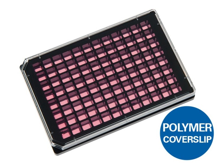
96 Well Plate | Black Walls & Flat Coverslip Bottom | ibidi

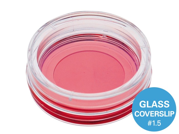
VWR®, Confocal Dishes for Microscopy | VWR

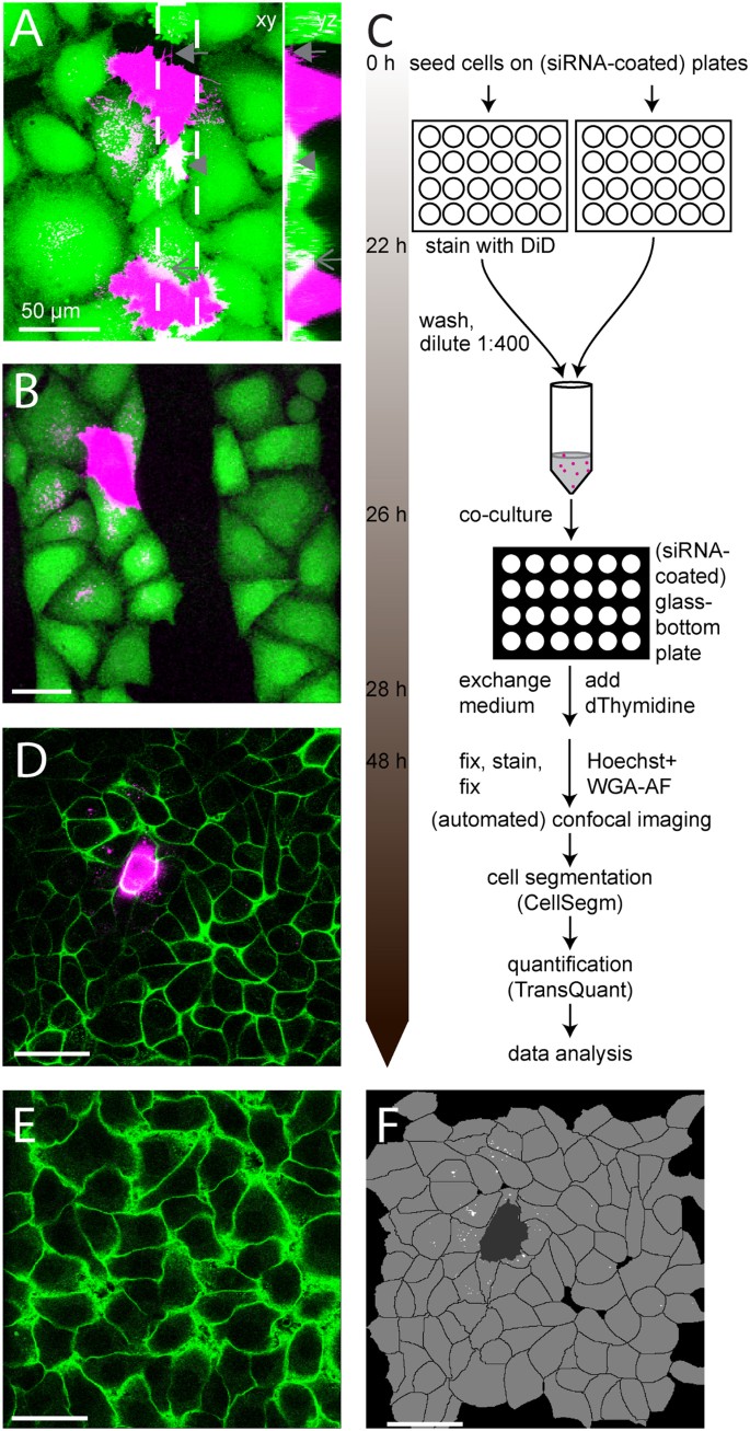
Novel microscopy-based screening method reveals regulators of  contact-dependent intercellular transfer | Scientific Reports
Novel microscopy-based screening method reveals regulators of contact-dependent intercellular transfer | Scientific Reports

NEST Scientific glass bottom cell culture plates and dishes from nest  scientific
NEST Scientific glass bottom cell culture plates and dishes from nest scientific

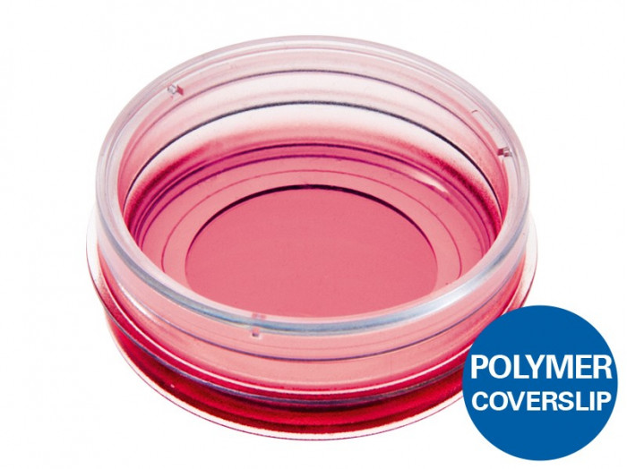
µ-Dish 35 mm, high | Cell Culture Imaging Dish | ibidi

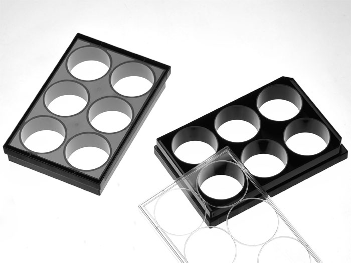
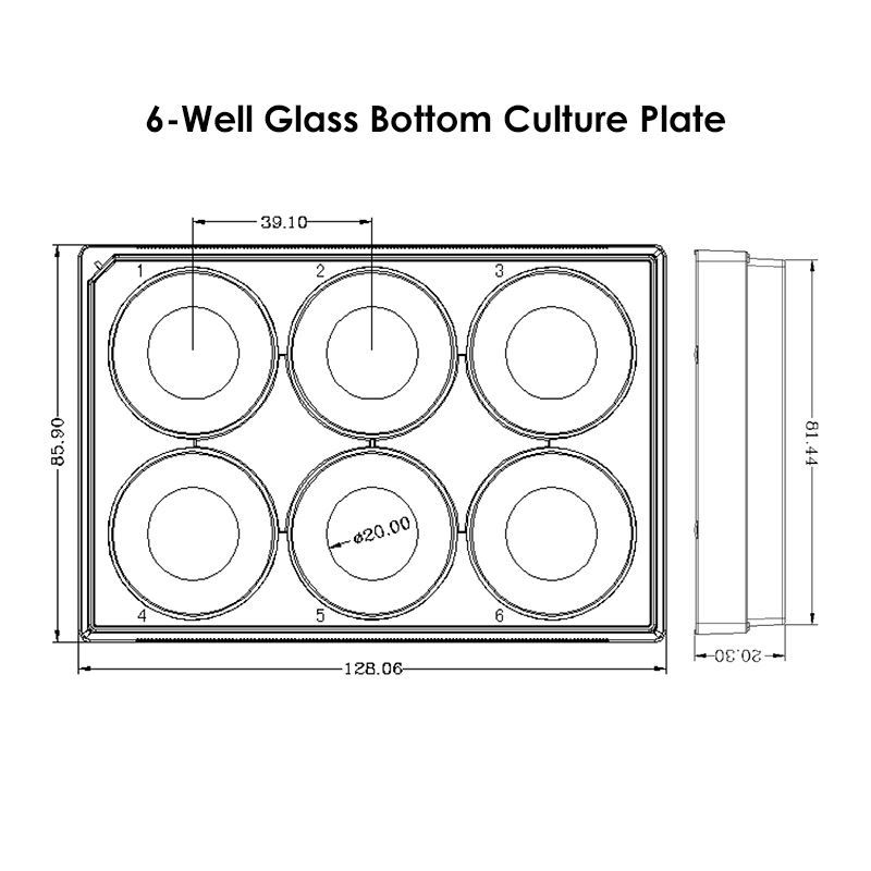
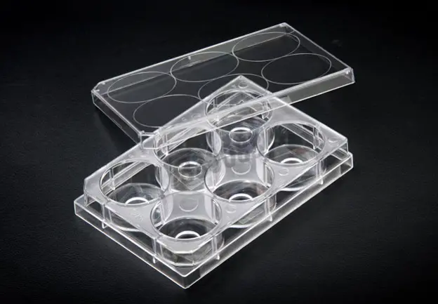
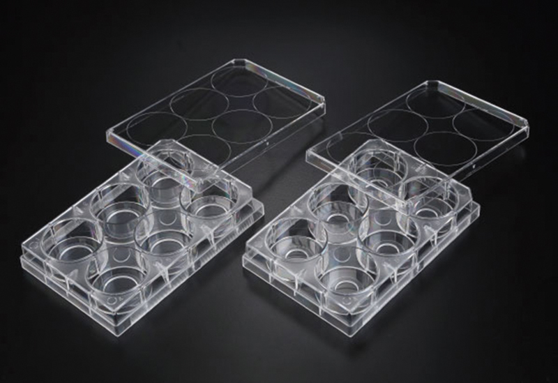
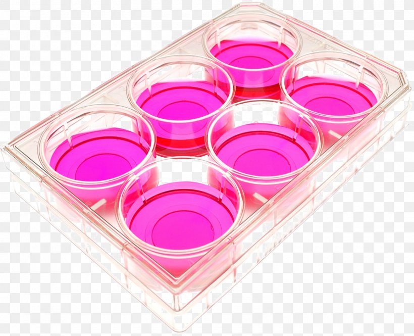
Sterile 6 Well Glass Bottom Cell Culture Plate - Glass Dia. 20mm:  Amazon.com: Industrial & Scientific
Sterile 6 Well Glass Bottom Cell Culture Plate - Glass Dia. 20mm: Amazon.com: Industrial & Scientific
Tech Tip: Imaging Bacteria Using Agarose Pads - Biotium

6 Well glass bottom plates | Cellvis

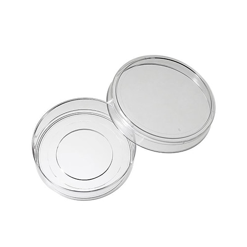
Glass Bottom Dish 35 mm | Live Cell Imaging Dish | ibidi

CLS-1812 - GLASS BOTTOM CELL CULTURE PLATES, TC TREATED, NEST- Chemglass  Life Sciences
CLS-1812 - GLASS BOTTOM CELL CULTURE PLATES, TC TREATED, NEST- Chemglass Life Sciences

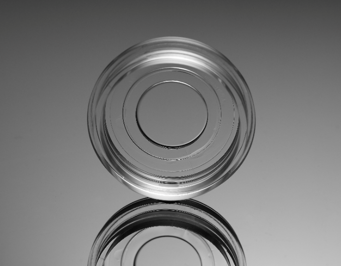
20mm glass bottom cell culture dish China Manufacturer

SPL Life Sciences
SPL Life Sciences

SPL culture plate for confocal microscopy - confocal region 1,33 cm² -  single bag -
SPL culture plate for confocal microscopy - confocal region 1,33 cm² - single bag -

12 Well plate with #1.5 glass-like polymer coverslip bottom, tissue culture  treated for better cell attachment than cover glass | Cellvis
12 Well plate with #1.5 glass-like polymer coverslip bottom, tissue culture treated for better cell attachment than cover glass | Cellvis

SPL Confocal Dish & Plate, Clear and Black, Glass and FLux, Adhesion and  Insert Type(id:9620064) Product details - View SPL Confocal Dish & Plate,  Clear and Black, Glass and FLux, Adhesion and
SPL Confocal Dish & Plate, Clear and Black, Glass and FLux, Adhesion and Insert Type(id:9620064) Product details - View SPL Confocal Dish & Plate, Clear and Black, Glass and FLux, Adhesion and
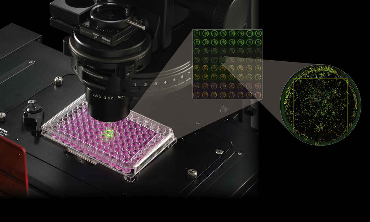
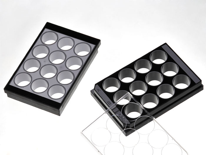
High Throughput Imaging | Life Sciences | Solutions | Nikon Instruments Inc.

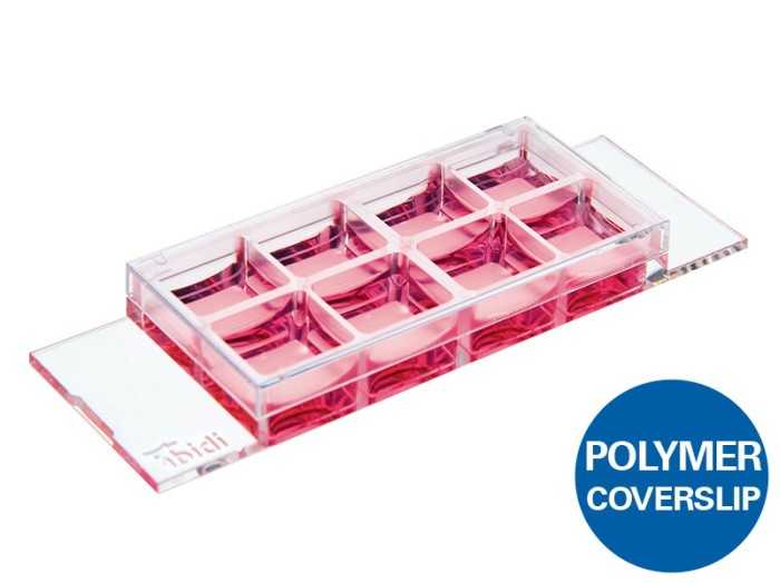
µ-Slide 8 Well | Chambered Coverslip for Cell Imaging | ibidi

SPL culture plates for confocal microscopy - SPL cell culture - Cell culture  - Serums - Media -
SPL culture plates for confocal microscopy - SPL cell culture - Cell culture - Serums - Media -

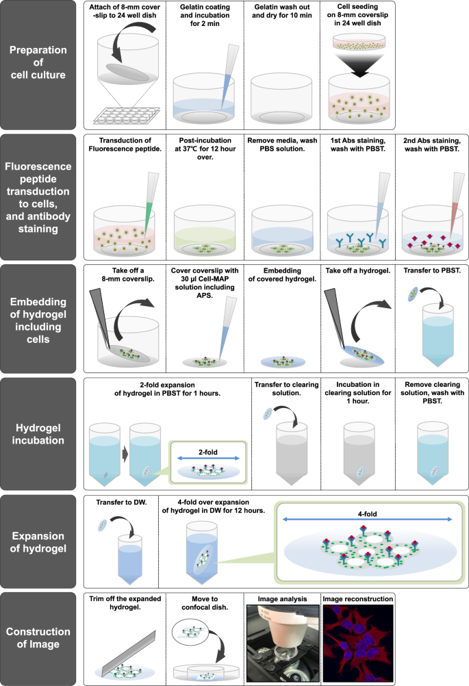
A Modified Magnified Analysis of Proteome (MAP) Method for Super-Resolution  Cell Imaging that Retains Fluorescence | Scientific Reports
A Modified Magnified Analysis of Proteome (MAP) Method for Super-Resolution Cell Imaging that Retains Fluorescence | Scientific Reports
Microscopy Solutions for Cell Culture

Confocal Imaging Dishes, Plates | Bioimager

Cell Culture Cell And Tissue Culture Petri Dishes, PNG, 1600x1298px, Cell  Culture, Cell, Cell And Tissue
Cell Culture Cell And Tissue Culture Petri Dishes, PNG, 1600x1298px, Cell Culture, Cell, Cell And Tissue

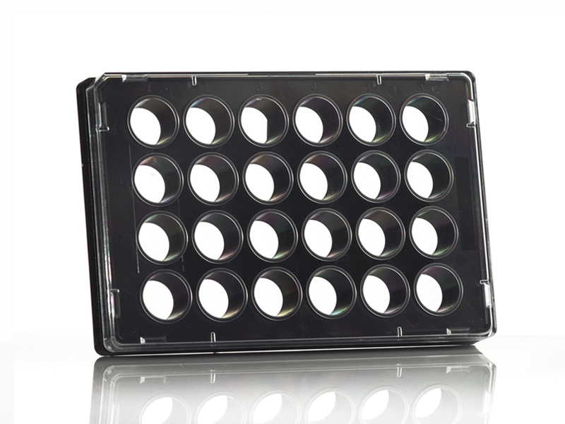
Tissue Culture Plate 24 Well, Clear Base | Azenta Life Sciences

PrimeSurface® 3D Culture Spheroid plates: Ultra-low Attachment (ULA) Plates  - S-Bio
PrimeSurface® 3D Culture Spheroid plates: Ultra-low Attachment (ULA) Plates - S-Bio

Immunostaining of lymphocytes analyzed by fluorescence and confocal... |  Download Scientific Diagram
Immunostaining of lymphocytes analyzed by fluorescence and confocal... | Download Scientific Diagram